What does an x-ray show?
- posted: Feb. 10, 2022
Radiographs (x-rays) are the first step in imaging of the injured or painful body part. Your provider can show you the x-ray at the time of your visit, and explain the degenerative changes in your joint, or identify where the fracture is and its severity.
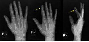
Hand X-ray – we take at least two images and sometimes add a third image at an oblique angle to properly show the body part. For example in this x-ray series of the hand, the injury is not visualized in the first AP image (left) but is shown in the next two images, oblique (middle), lateral (right). This shows a dislocated finger. For hand and wrist conditions, schedule an appointment with our upper extremity specialists.
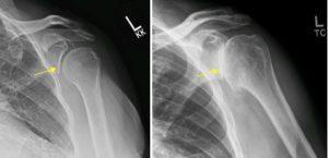
Shoulder X-ray – An x-ray can give us a lot of information about what is causing your joint pain. The x-ray on the left still has space in the glenoid cavity (shoulder joint) and the one on the right has significant osteoarthritis, with bone on bone. This condition can be very painful and debilitating. A shoulder joint replacement surgery may be your best option. Schedule an appointment with one of our shoulder doctors to learn about your options.
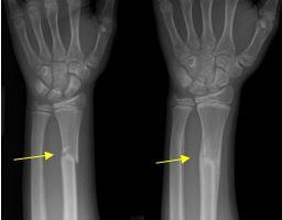
Wrist X-ray – Fracture Care – These two images show the healing process in a wrist fracture. The left image was the initial image taken when the injury occurred. The wrist was casted and the progress of healing was monitored as the body lays down new bone at the fracture site. The right image was taken 10 weeks after the injury, showing how the bone is healing in the remodeling phase.
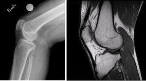
Knee X-ray vs. MRI – On the left is a lateral (side) view of a knee and on the right is the same view using MRI, Magnetic Resonance Imaging. X-ray clearly visualizes the bone and alignment and can rule out any abnormal growths, degeneration, fractures or dislocation. MRI clearly visualizes the soft tissue (muscle, ligament, tendon, fluid) and integrity of the joint. To learn about your treatment options, please schedule an appointment with one of our knee doctors.
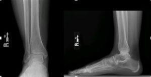
Foot & Ankle X-rays – Weight bearing imaging can be used as a tool for the providers to see instability and joint space alignment in the ankle that a non-weight bearing x-ray may not identify. We have a Foot & Ankle Specialty team that can work with you to find the best treatment option.
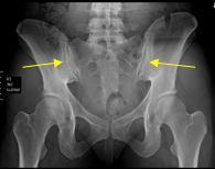
SI Joints – The Sacroiliac (SI) Joints are the major connection joints between the upper and lower body. It is located between the ilium in the pelvis and the sacrum at the base of your the spine. If this joint is involved in a trauma, like a car accident it can cause pain that starts in the lower back and buttock and may radiate to the hip, groin or upper thigh. Dr. Jeffrey LaPorte has a special interest in the dysfunction of the SI joint.
The post What does an x-ray show? appeared first on Missoula Bone & Joint.